


BioREE-B™ is a lanthanoid staining technology for the study of live, unfixed cells
Highly informative SEM images without complex sample preparation


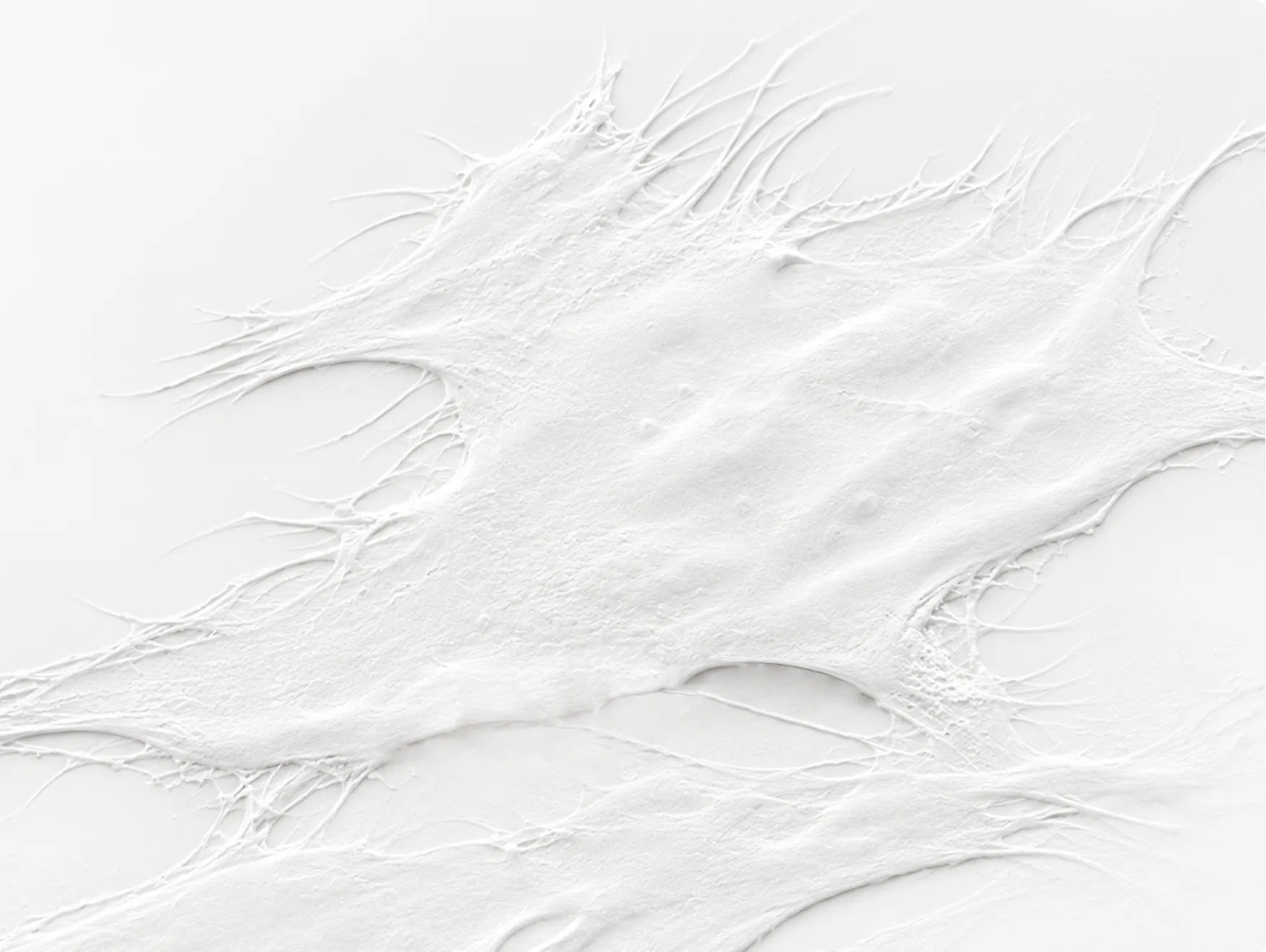
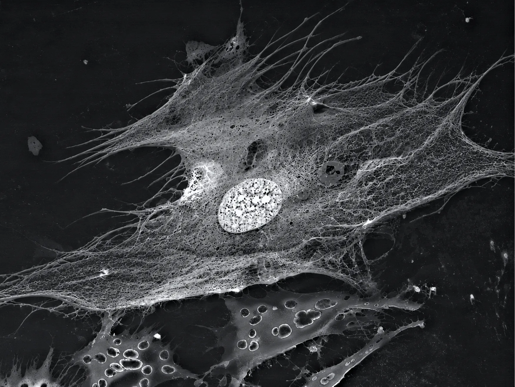
A fundamentally new sample-preparation method is now available as a practical laboratory tool
BioREE-B is a reagent kit for two-stage supravital staining of biological samples. It enables the surface, the immediate subsurface layer, and elements of internal cellular structure to be examined without prior fixation, dehydration, or conductive coating.

Reduces the time and number ofpreparation steps

Eliminates the need for prior chemical fixation in compatible applications
Supports work with samples that are sensitive to conventional preparation

Tested by ZEISS Research Microscopy Solutions
The analysis of prestained cells was possible in the FESEM without additional conduction or coating of the sample.

ZEISS Research Microscopy Solutions, report author
Preservation after storage. After several days, some single bacteria with well-preserved ultrastructure could still be observed.
Ease of handling. The report describes the staining procedure as apparently easy to handle.
BioREE bridges fundamental science and solutions for multiple industries
We have turned an innovation into a practical tool
BioREE-B adds visual morphological data to quantitative methods and supports research into cells, microorganisms, and their interactions with materials
Pharmaceuticals and biotechnology

Microbiology and cytology

Medical devices and biomaterials

Ophthalmology

Food industry and agricultural biotechnology

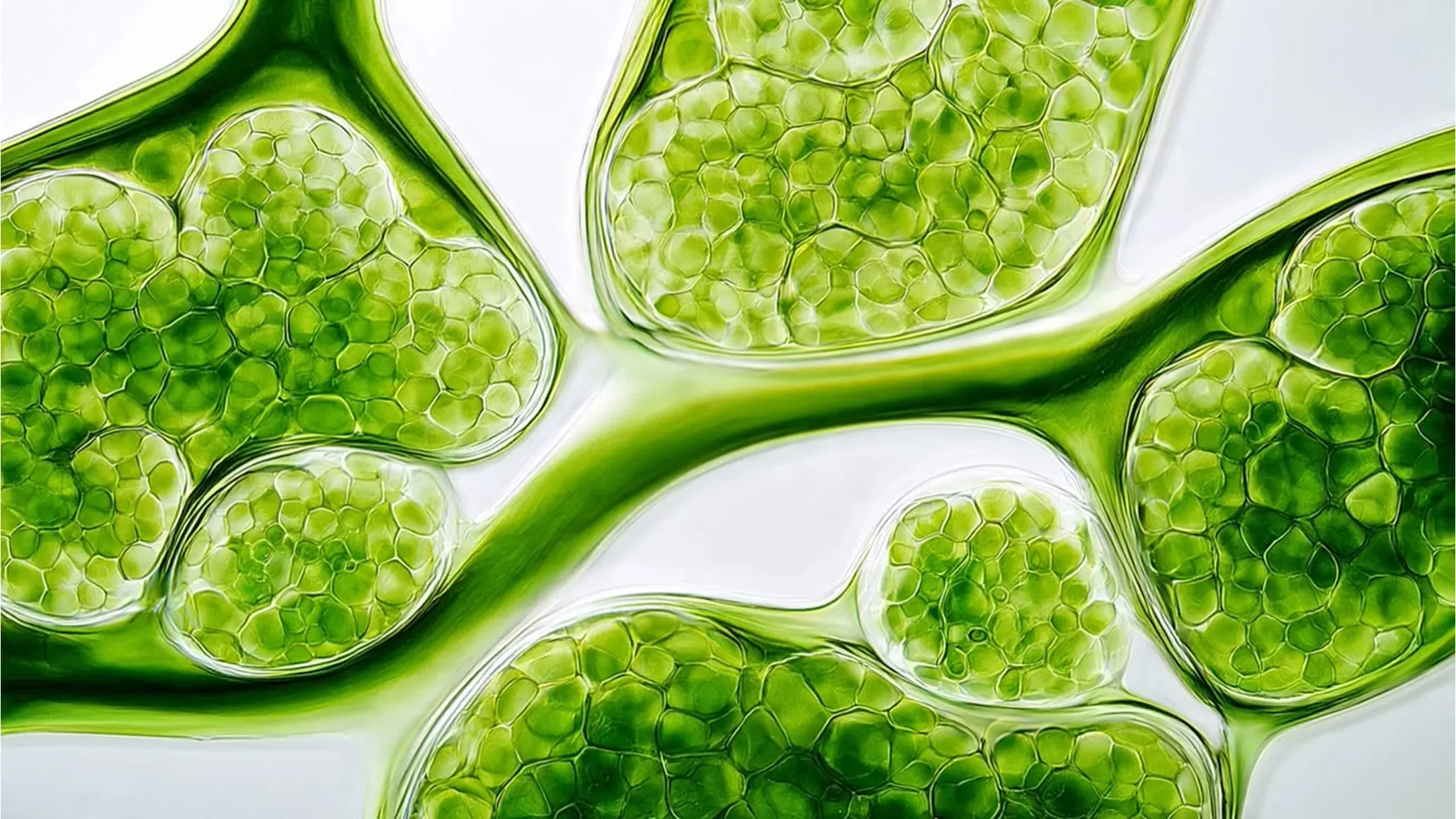
Broad applicability — from cell cultures to surgical specimens
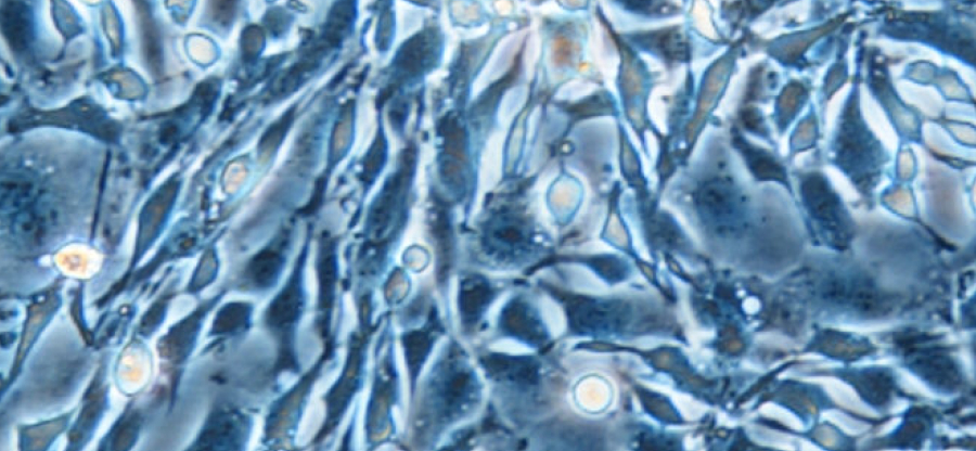
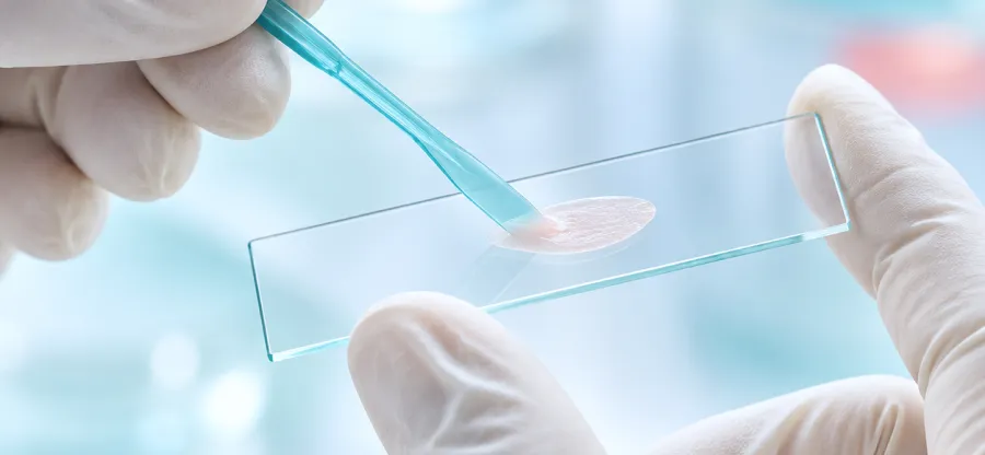
samples
Equipment requirements: A scanning electron microscope with a low-vacuum or variable-pressure mode and a backscattered-electron (BSE) detector. Operating parameters are selected according to the microscope model and sample type.
Discuss applications →Conditions and limitations
• The method is not intended for previously fixed tissue blocks.
• Phosphate-based substrates are not recommended; bone and teeth are also unsuitable for examination.
• The sample support must withstand mildly acidic conditions.
Reduce the number of preparation steps and obtain more structural information
BioREE-B provides an additional research mode for applications where minimal sample deformation and richer morphological information are important.
BioREE-B provides an additional research mode for applications where minimal sample deformation and richer morphological information are important.
Two-stage staining of a live, unfixed sample
Examination of a compatible sample without prior coating
The absence of a coating preserves more of the sample’s original information for microanalysis
Surface, immediate subsurface layer, and elements of internal structure

Expand the capabilities of SEM for studying biological samples
Compared with conventional sample-preparation methods, BioREE-B extends the capabilities of SEM by supplementing surface images with information about structures in the immediate subsurface layer.
• High-resolution visualization of ultrastructure in the immediate subsurface layer.
• Identification of cells on complex objects and substrates through enhanced contrast.
• Comparative information about the functional state of cells within a single sample.
• More accurate chemical microanalysis without prior conductive coating.
The method is not a stand-alone quantitative assay of metabolism, a method for species-level identification of microorganisms, or a clinical diagnostic test.
Sequential treatment with BioREE-B enhances compositional contrast in biological structures and expands the range of elements that can be distinguished in SEM images.
Sequential treatment with BioREE-B enhances compositional contrast in biological structures and expands the range of elements that can be distinguished in SEM images.
Two-stage staining makes more structures available for analysis

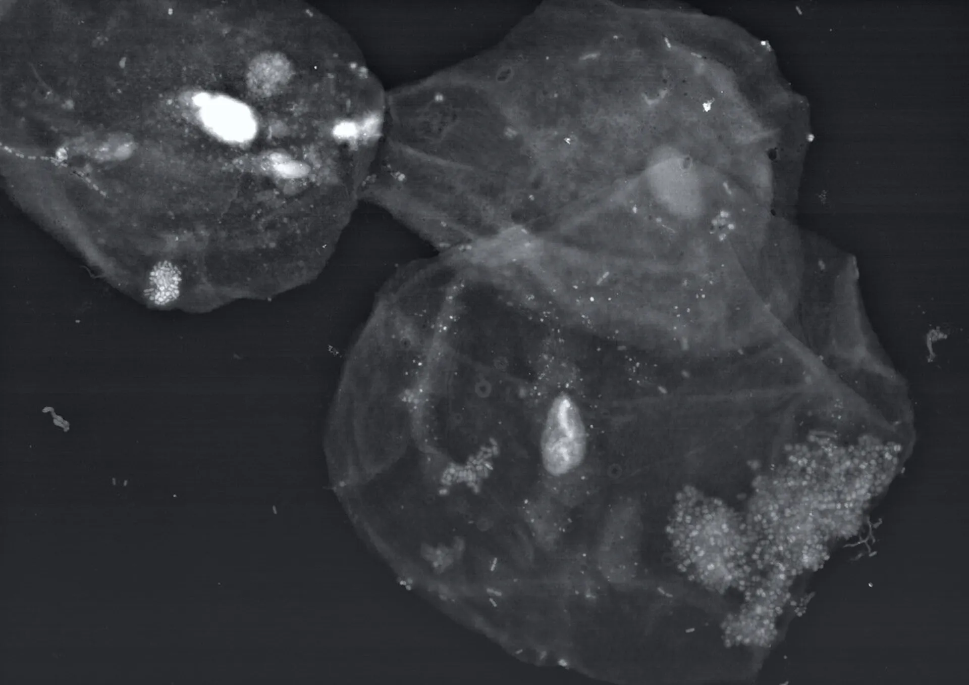
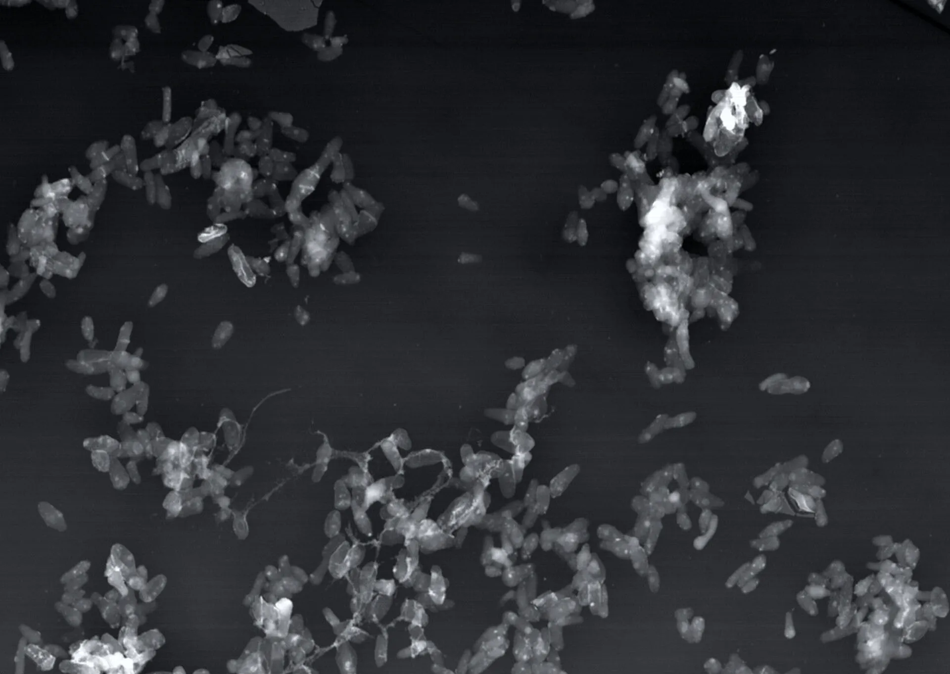
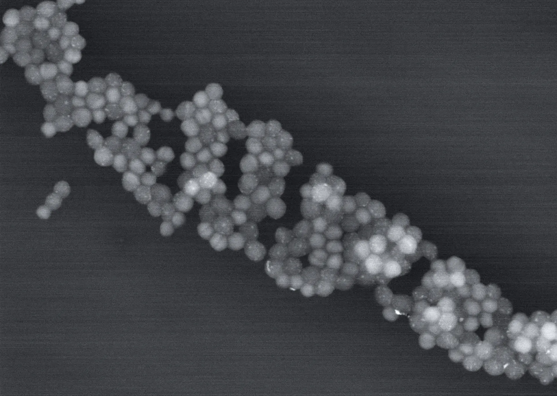
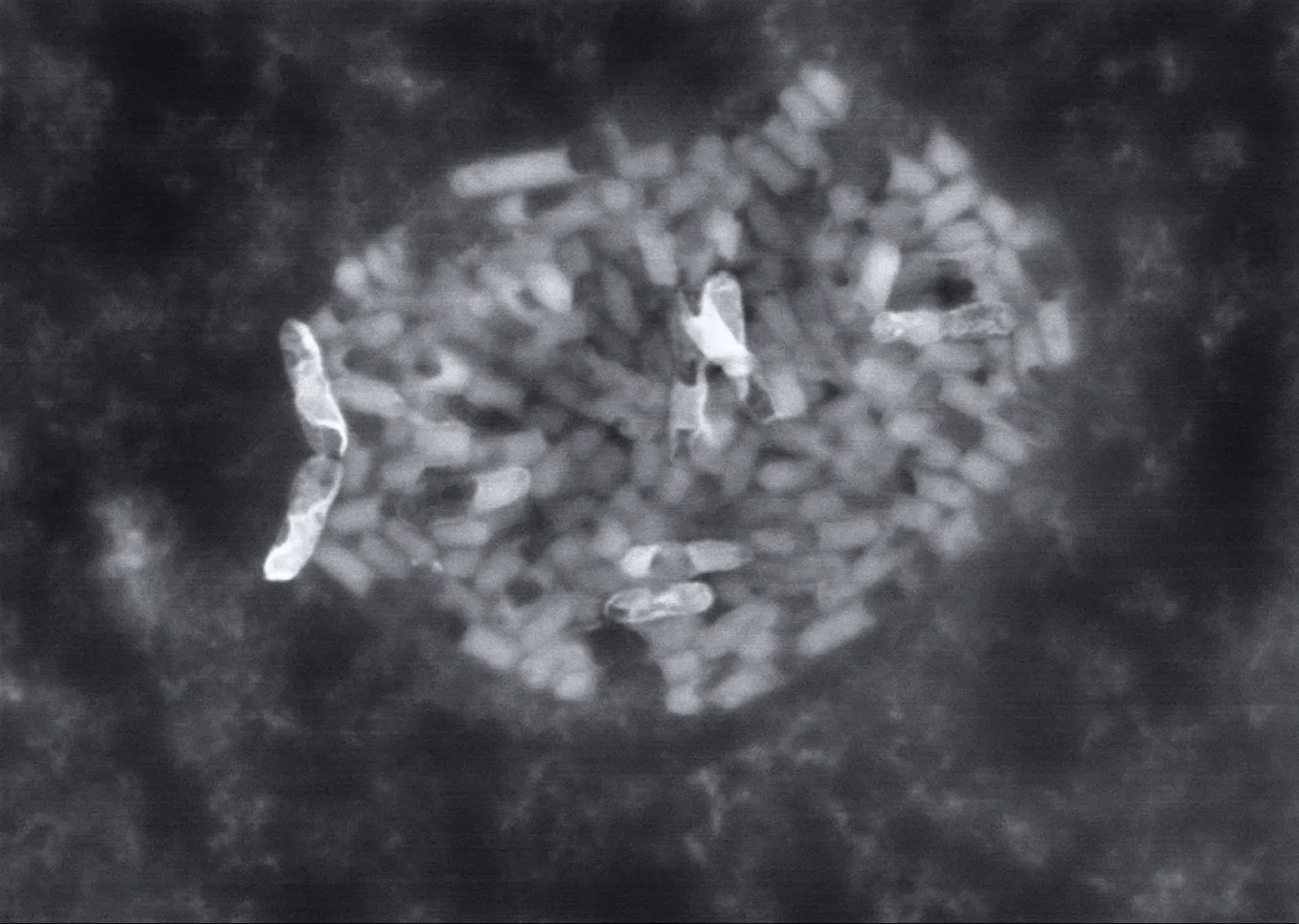
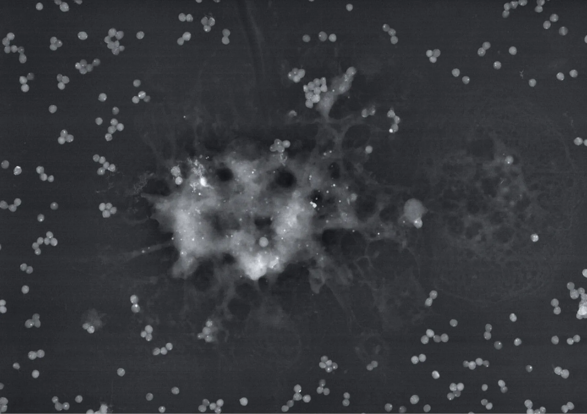
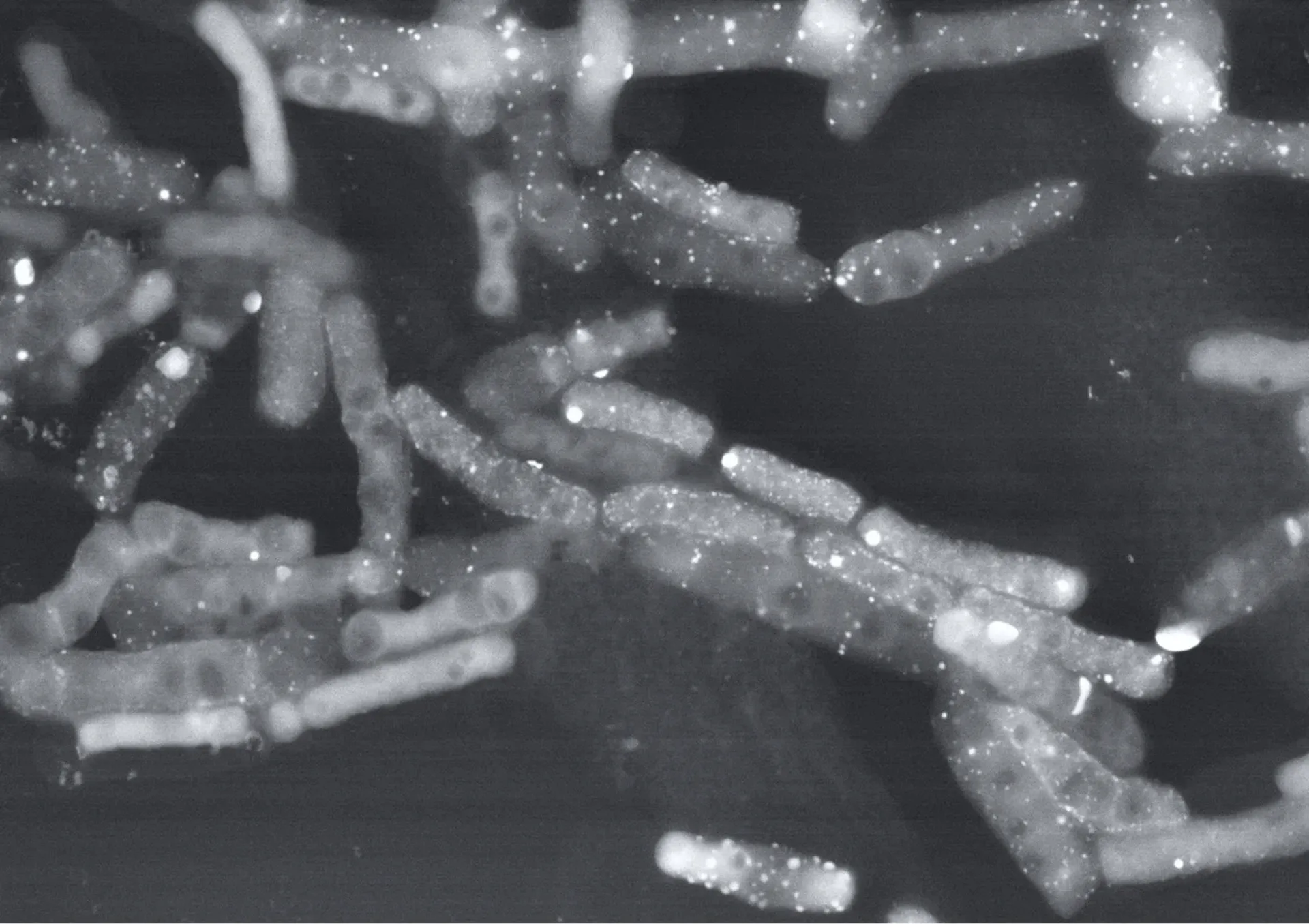
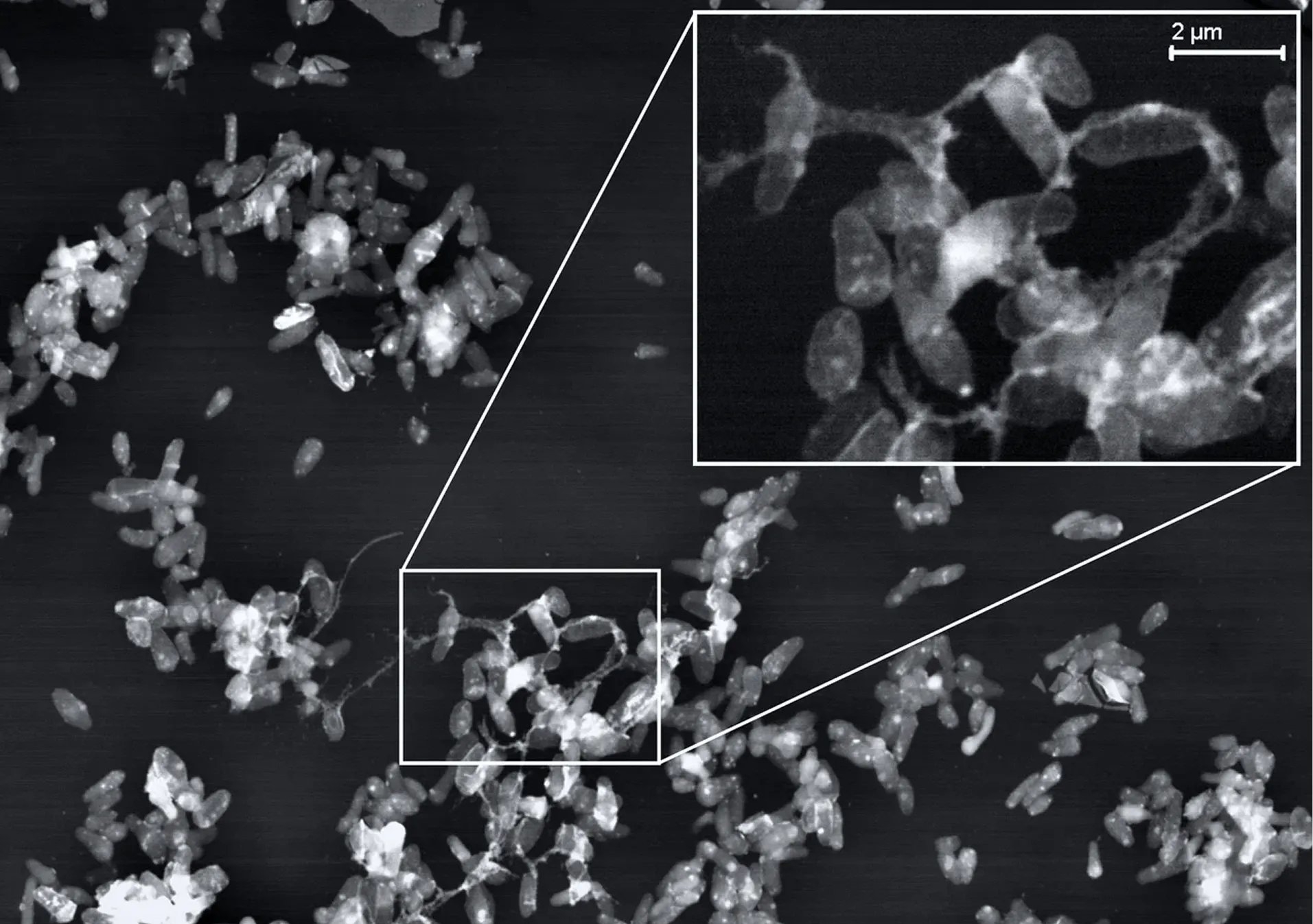
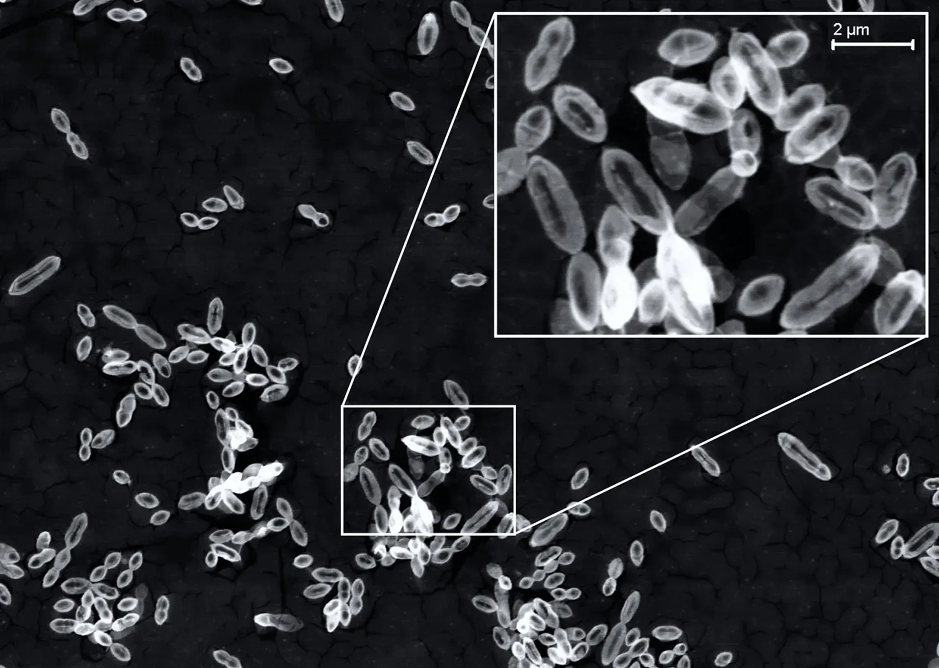
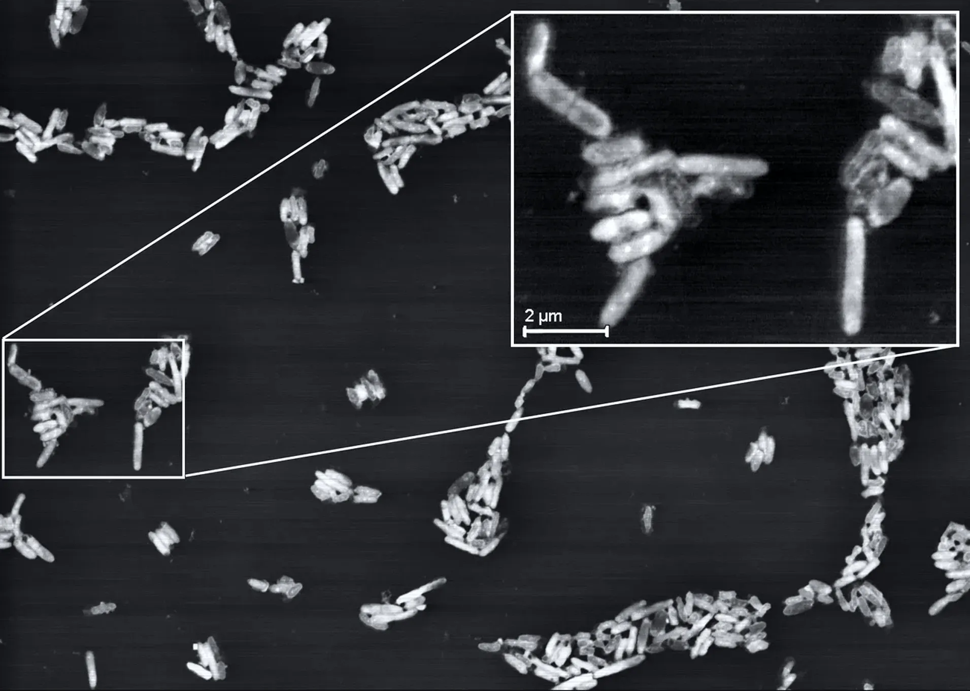
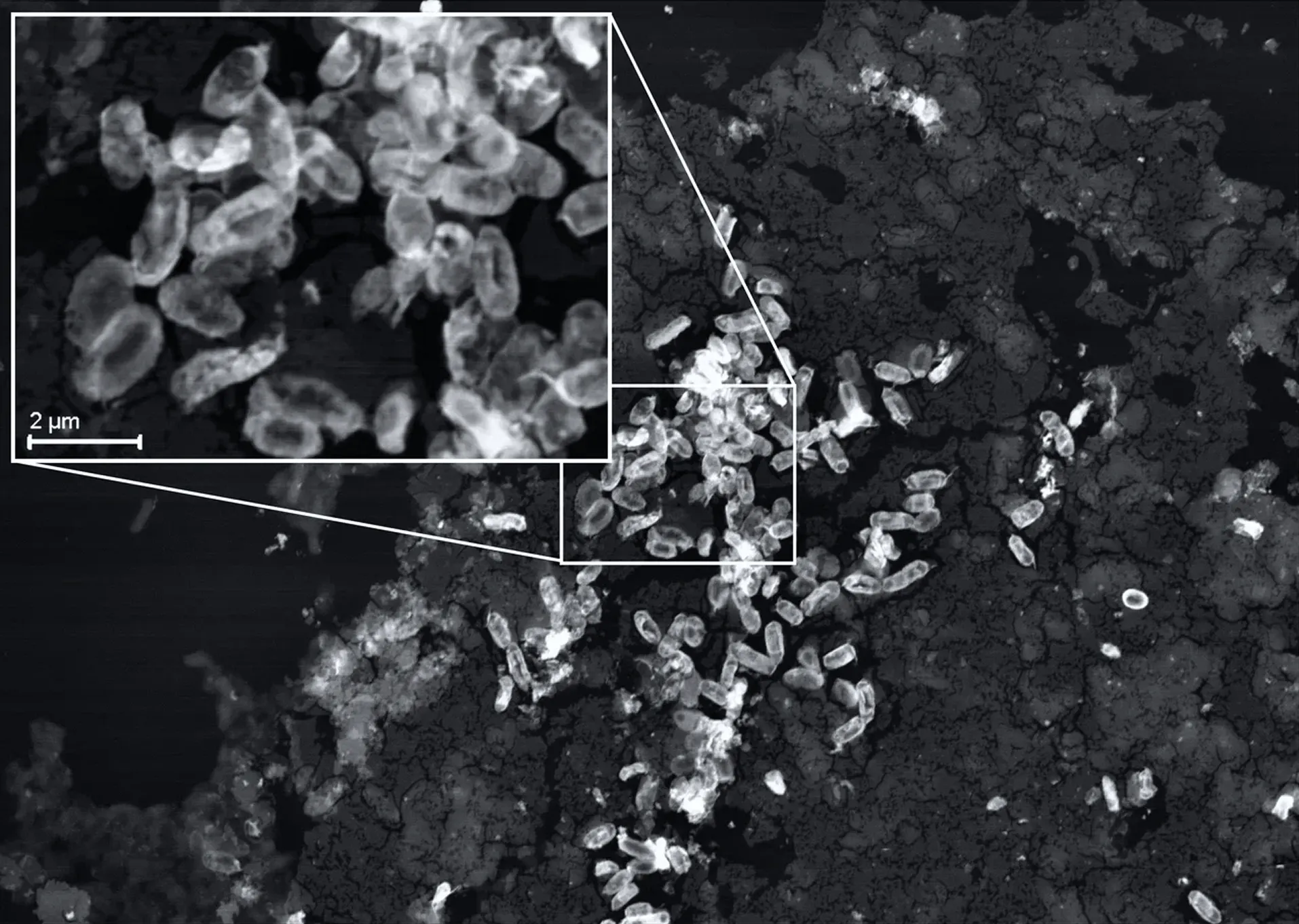
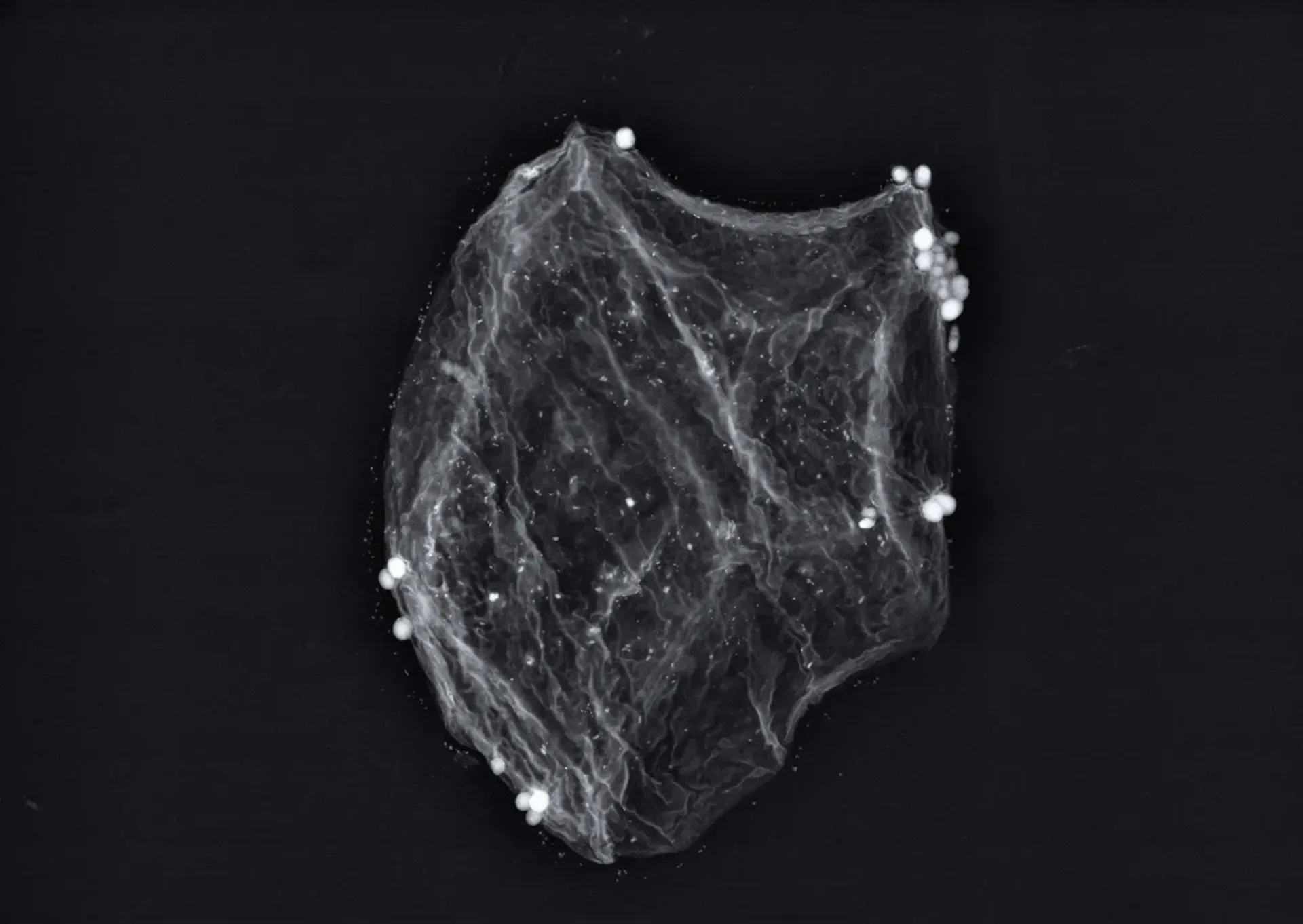
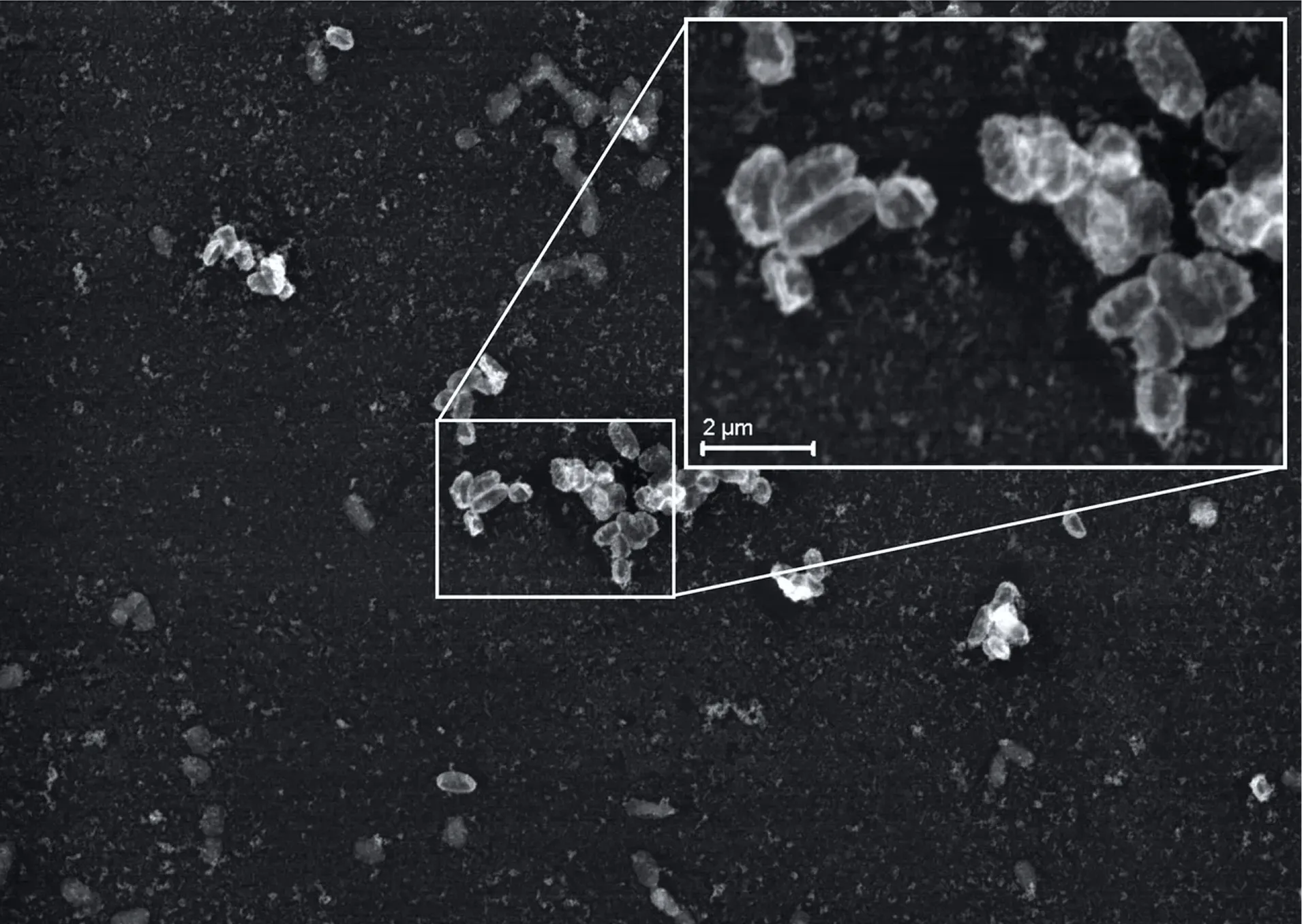
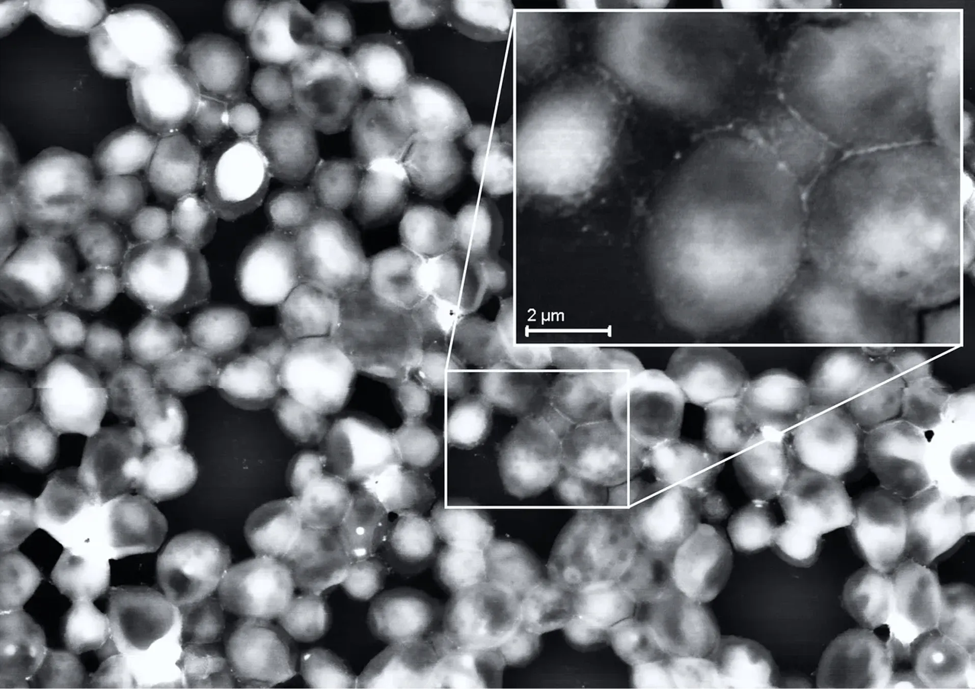
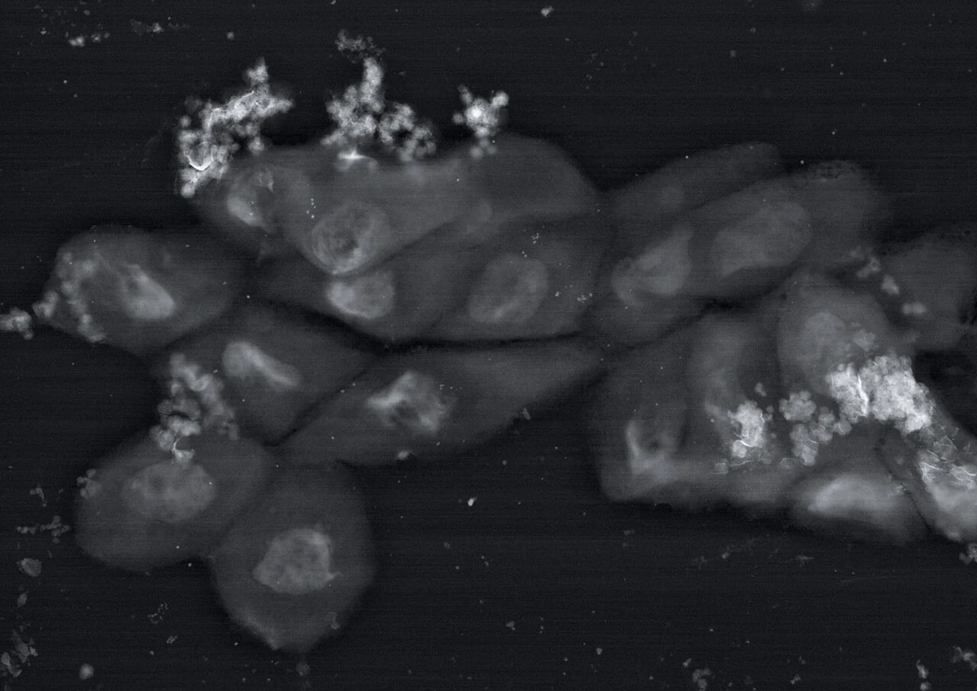
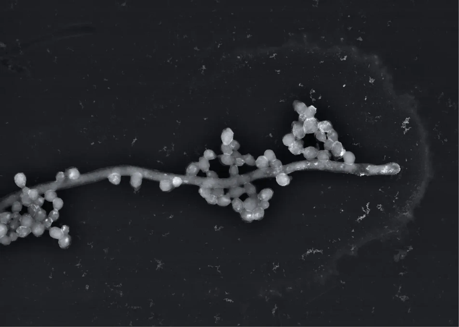
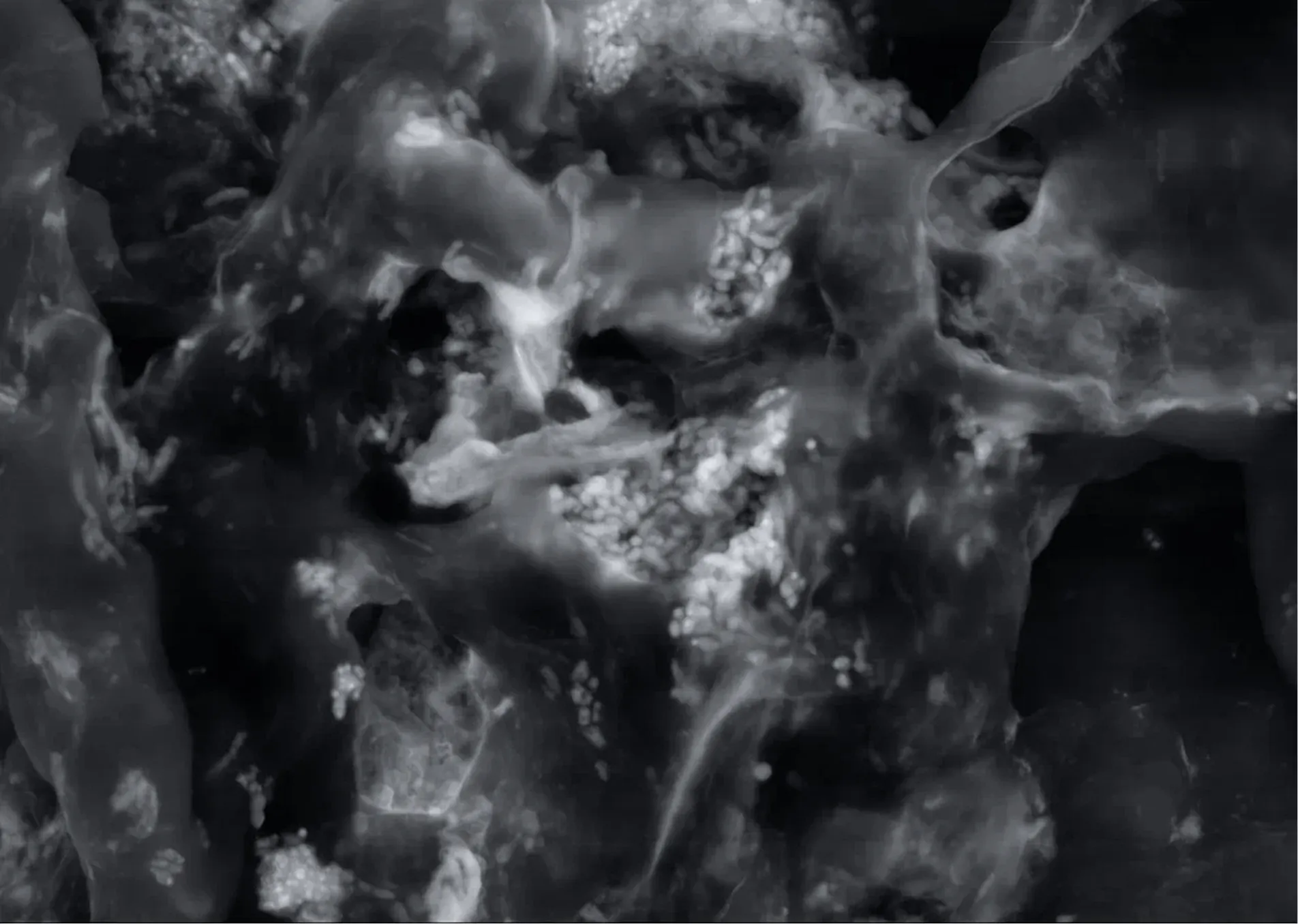
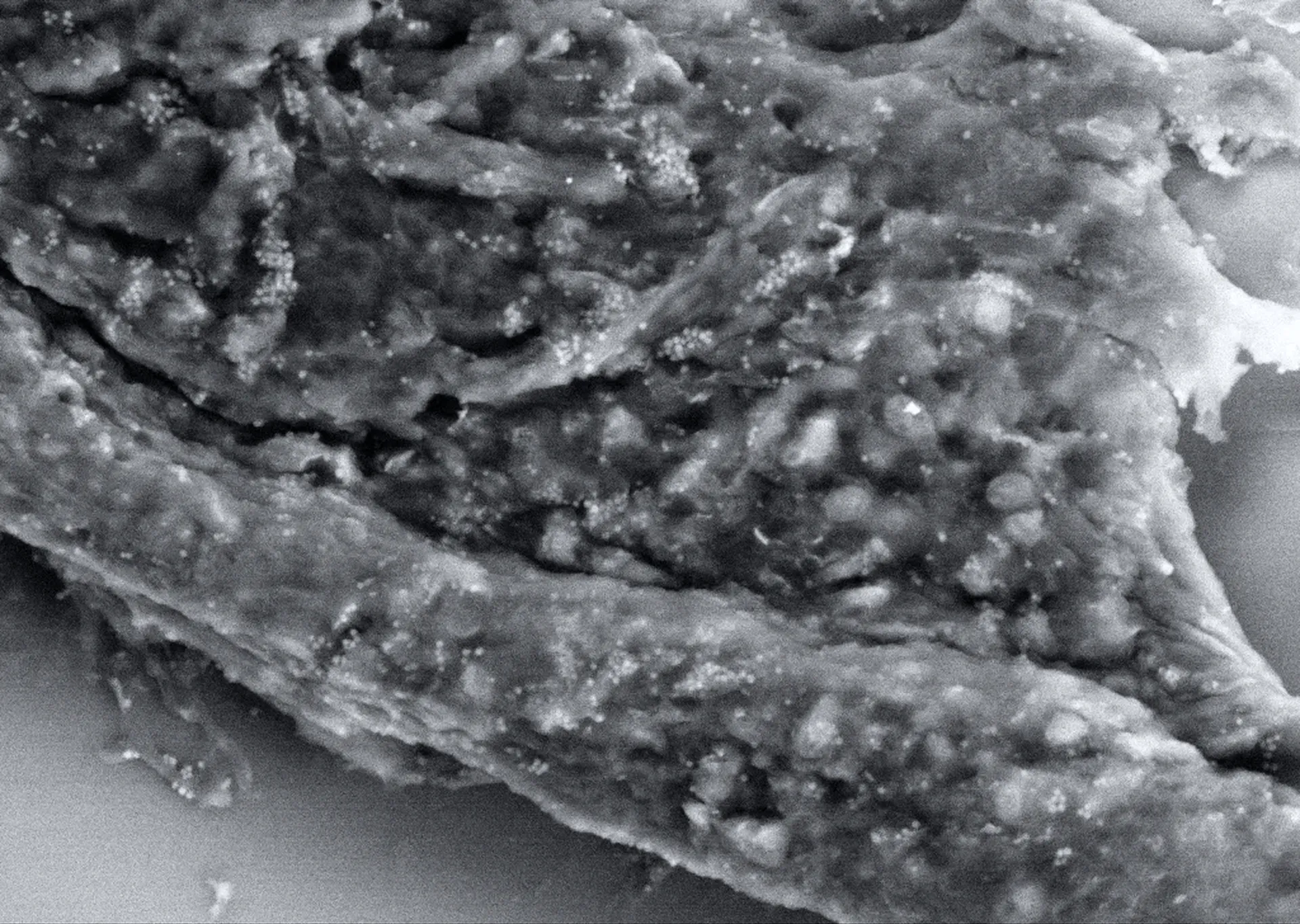
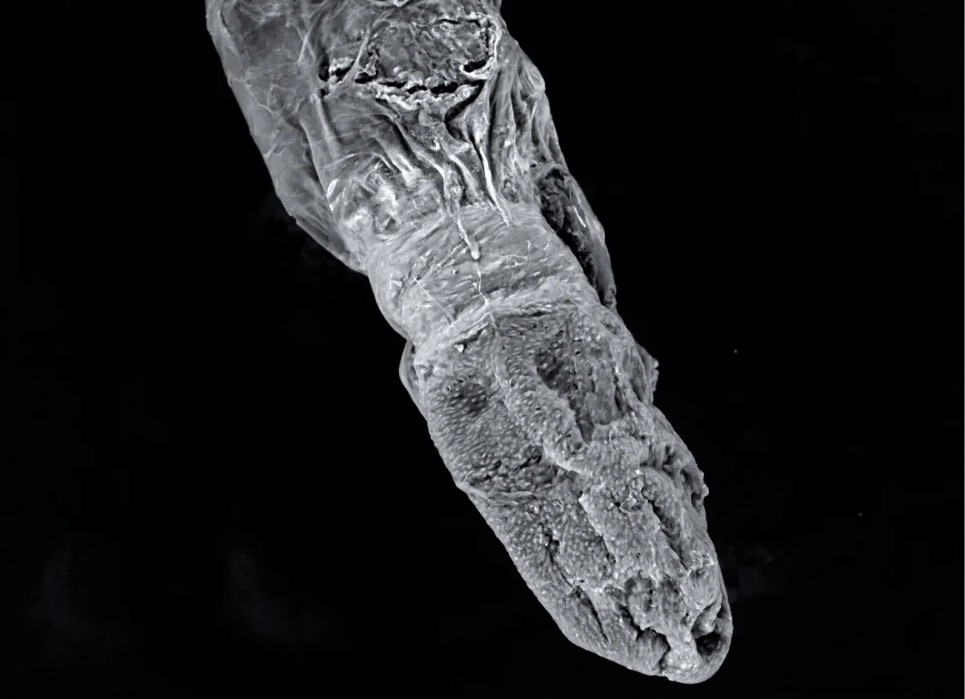
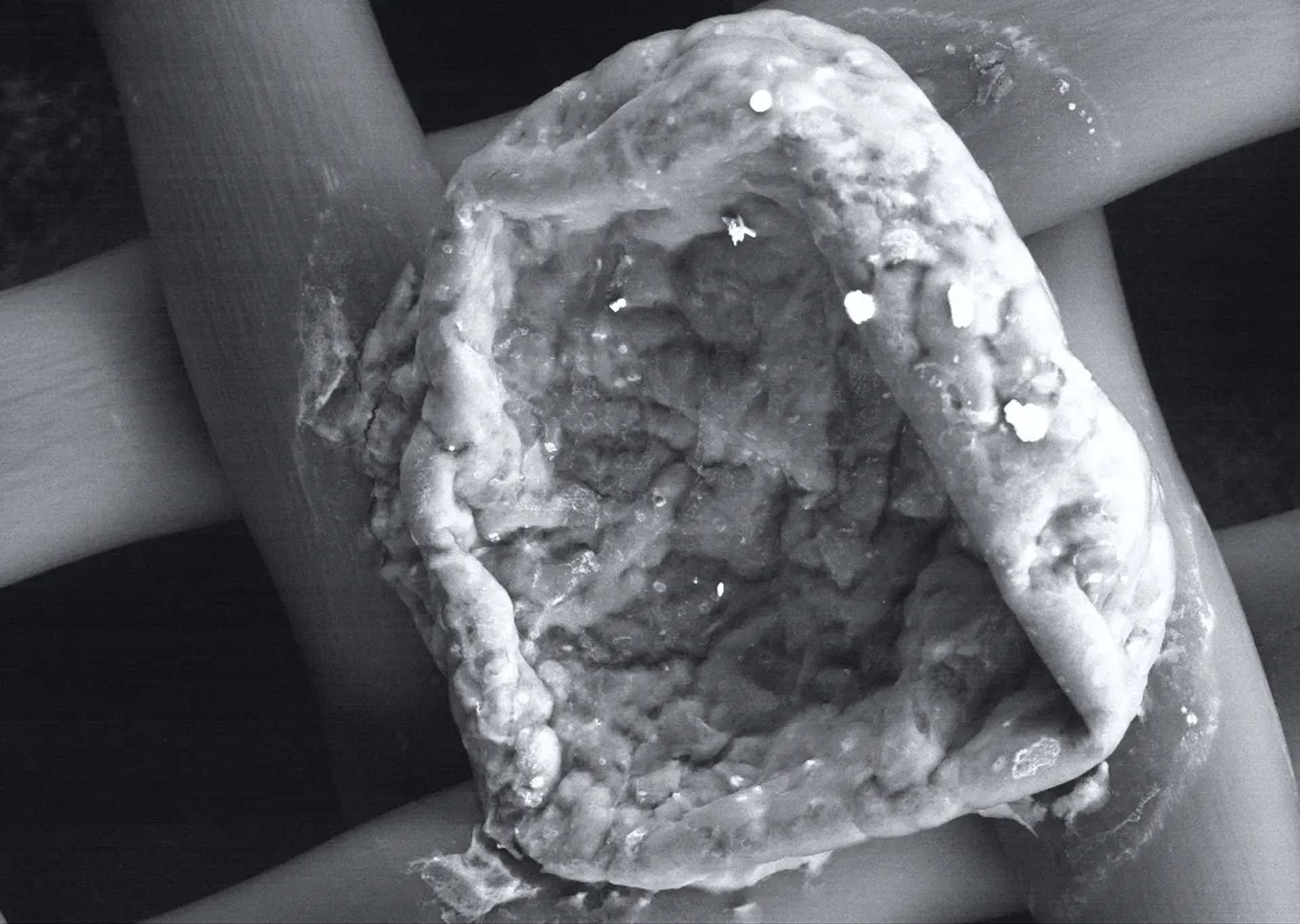


Explore gallery


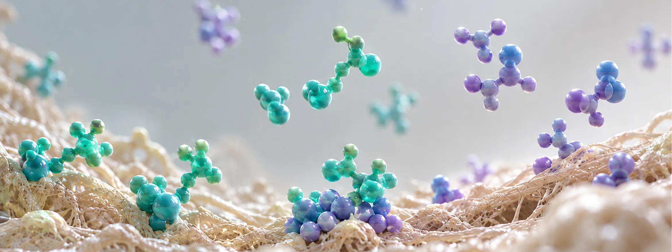
Scientific principle of the technology
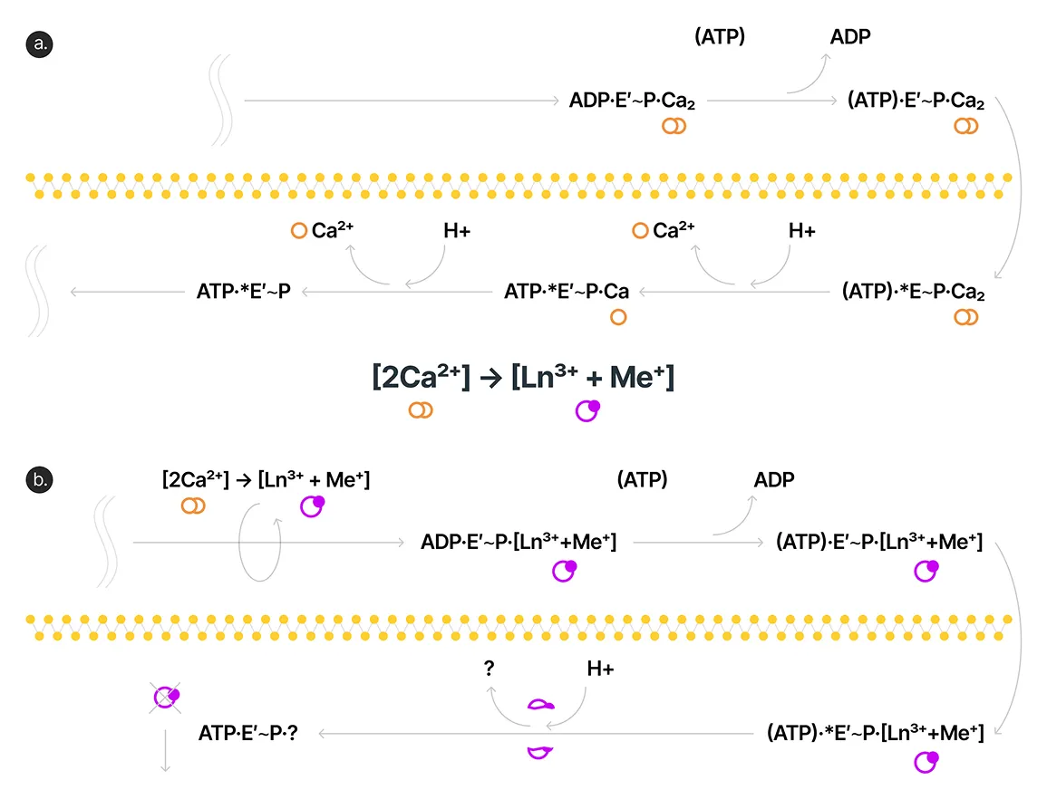
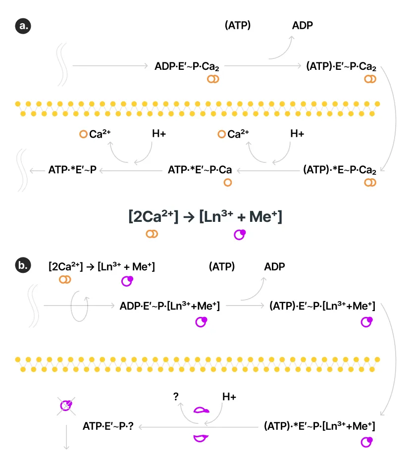
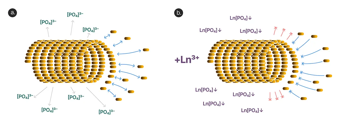
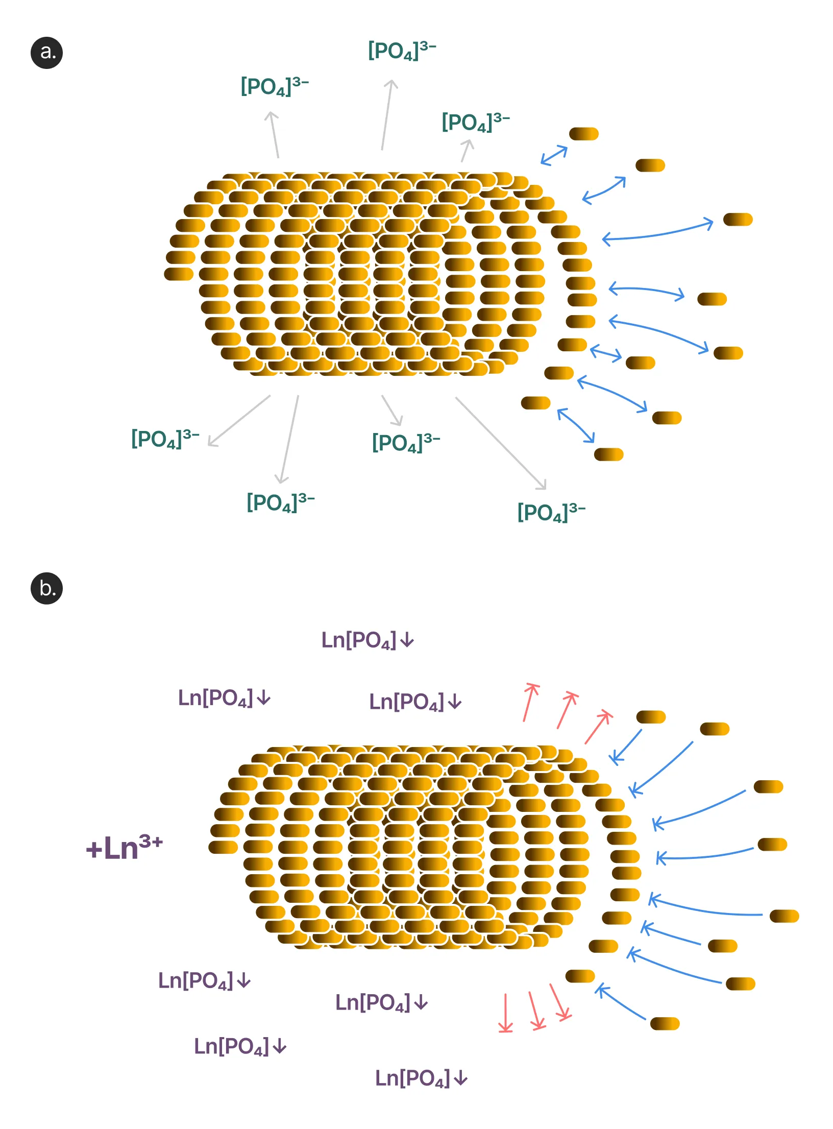
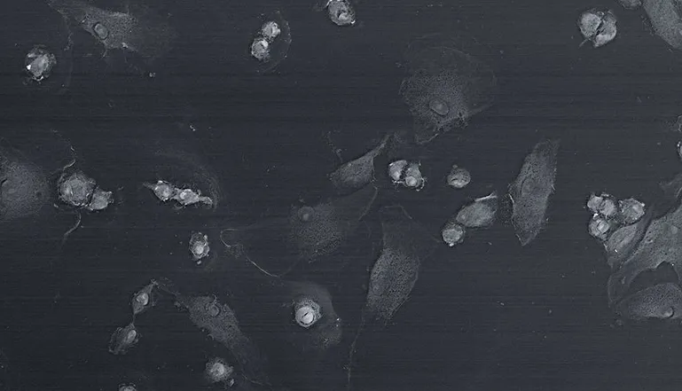
BioREE-B combines lanthanoid staining with an additional treatment using lead salts. A live, unfixed sample is treated sequentially with two formulations that selectively label different biological structures.
As a result, the surface, the immediate subsurface layer, and selected elements of internal structure become accessible for examination in SEM images.
Staining also stabilizes the cells’ natural dehydration and helps reduce sample deformation during examination.
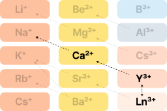
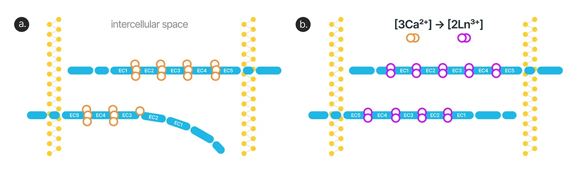
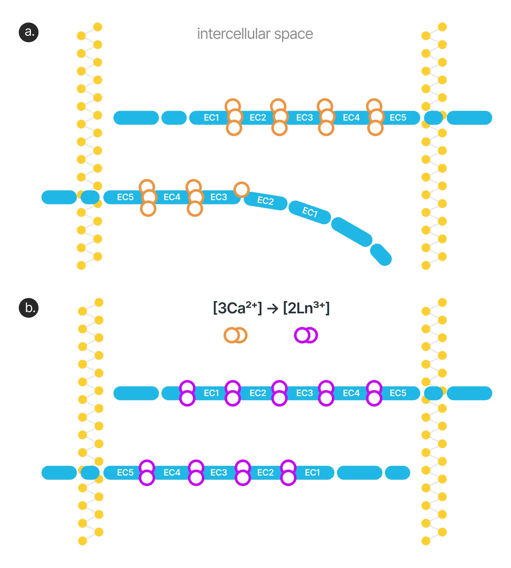
.webp)
.webp)



Explore mechanism
A sequential workflow — from sample preparation to SEM imaging
Compatibility
Preparation
Staining

Final Preparation
SEM Imaging



Protocol for use
The technology is described inscientific publications, protected by a patent, and tested on SEM equipment
Fast visualization of ocular surface microbiota

Rapid whole-cell sample preparation for SEM
Reversible metabolic arrest with lanthanoids
Technical testing on the ZEISS GeminiSEM 300
Start using BioREE-B in your laboratory

Submit an inquiry to learn more about thetechnology, clarify equipment requirements, andreceive current BioREE-B supply terms



